Multi MUP Reference Limits
Enlarged database for MultiMUP Reference Limits
After extensive collection of reference data and statistical processing (see below), MultiMUP normal reference limits for the following muscles are now
available:
|
|
- Abd pollicis brev
- Biceps
- Bic fem cap brev
- Deltoideus post
- Ext dig brevis
- Ext dig communis
- Ext indicis
- Flex carpi rad
- Frontalis
- Gastroc caput med
- Gluteus medius
- Infraspinatus
- Inteross dors I
- Orbicularis oris
- Paravert L5
- Sphincter ani
- Splenius capitis
- Tibialis anterior
- Trapezius
- Triceps
- Vastus lateralis
|
|
MultiMUP Reference Limits is compatible with the following EMG systems:
- Keypoint®Classic
- For Keypoint®Net, see PowerPack
Usage
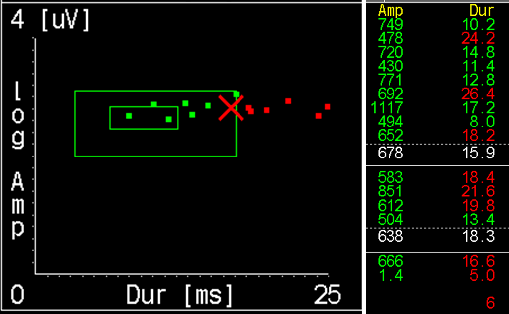
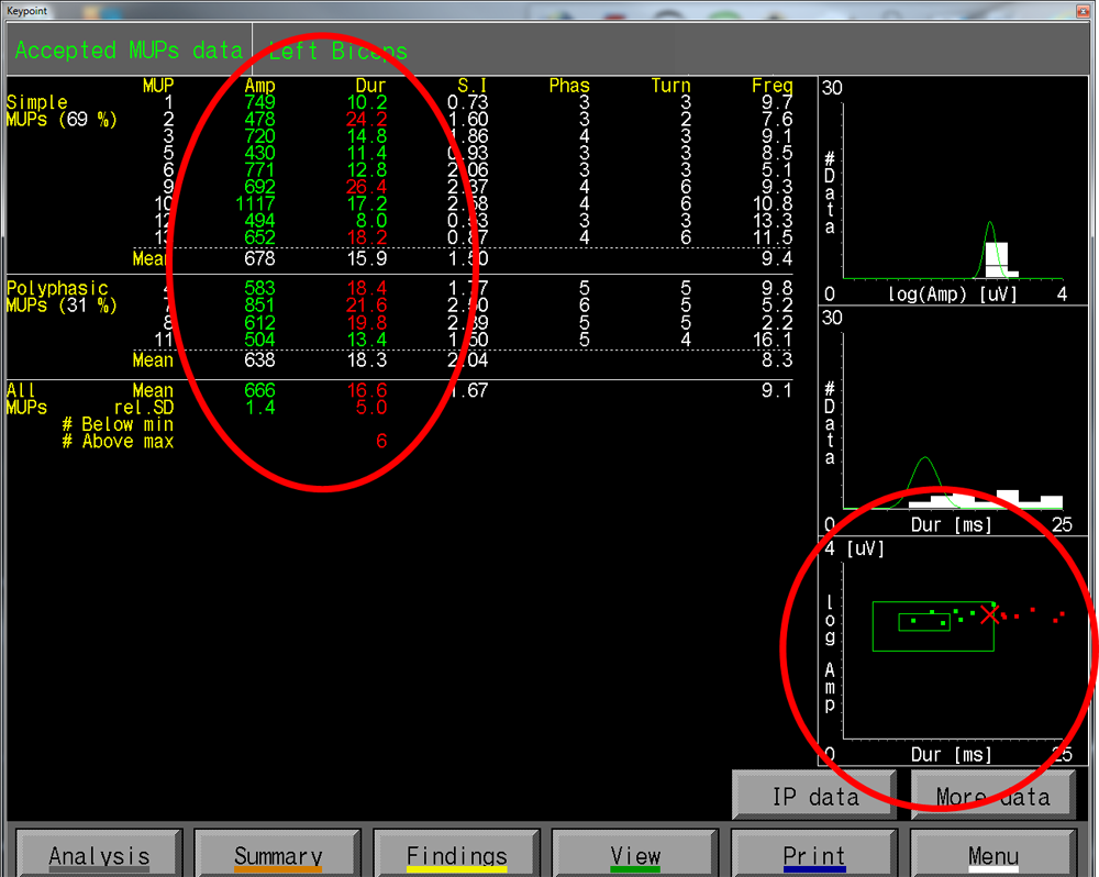
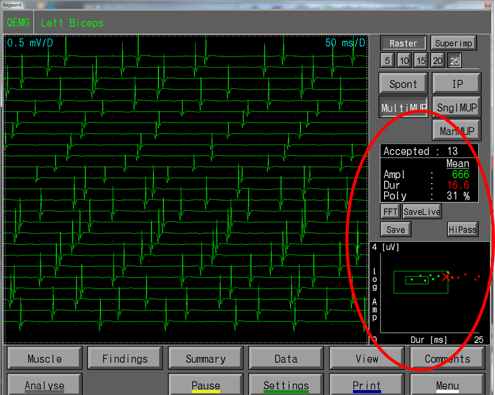
MUP amplitudes and durations are instantly compared with the normal reference limits during the examination.
Reference limits for both mean values and individual values are included.
Green numbers and dots in the Dur-Amp plot, indicate normal while red indicates an abnormal MUP.
The two green boxes in the dur-amp graph shows the normal borders for the mean result (inner green box), as well as limits for outliers (individual) values (outer green box).
Data Collection
Participants in collecting reference material
The collection of reference material for Multi-MUP analysis started in Uppsala
already when the method was established 1994(2). With increasing knowledge of
the technique and increasing demand of more reference material, many other
centers have contributed to the collection of data. The principle has been as
follows. When a new laboratory wanted to participate, the physicians from that
laboratory were asked to send values (via internet) from 10 healthy controls to
Uppsala for inspection. Often 30 MUPs from each subject was recorded, and
editing was performed in Uppsala with feed back comments. In other instances 20
edited MUPs were delivered for inspection.
Values from the 10 controls were then compared to the existing database created
in Uppsala. If no difference in any respect was detected, the laboratory was
“accepted” as partner and could start to collect reference material in the
muscles that were in focus at that time. Only one laboratory has shown a slight
deviation from the existing mean values. This has been discussed and possible
methodological differences have been minimized. Reference values from the
Sphincter Ani muscle, is taken from a collaborative study with Dr Podnar as main
contributor(4). The patients are all investigated at his laboratory.
Methods for MULTI MUP recording
Concentric needle electrodes have been used. The electrode is inserted
perpendicularly to the skin, a few mm beneath the fascia. The electrode is then
redirected in skew angels and with different depths. Two or three skin
insertions, spaced at least 10 mm, preferably perpendicularly to the fiber
direction have been performed.
A weak contraction has been used, usually giving 3-5 MUPs from each recording. A
special notice has been taken to a constant recording position during 5 seconds
preceding the analysis. 30 MUPs have been aimed for, to allow easy editing by
deletion, rather than by resetting cursors.
Control material
Selection
Normal subjects and healthy patients were included. Normal subjects were staff
members and visitors to the laboratories. Alternatively, patients, after
informed consent, were included if they were seen in the EMG lab for a focal
lesion, not affecting the investigated limb or segment.
Age
Age groups between 15 and 80 (occasionally higher) were included. A somewhat
uneven distribution is seen for some muscles as a result of practical factors.
Gender
No special effort has been made to recruit same number of men and women. In our
earlier and in this material, we have not seen any correlation between MUP
parameters and gender. A dominance for one gender or the other is therefore not
influencing material.
Statistical methods
Mean
Individual studies:
Mean values were calculated for each parameter, except for polyphasicity for
which the occurrence in % was given.
All data from each muscle:
The total material from each muscle was inspected regarding general distribution
and aberrant data. If individual data were detected that might influence the
material, this was treated according to the rules given elsewhere(3).
The influence of height, gender and age were analyzed by stepwise multiple
linear regression. Usually only age was influential, and also this occurred only
in a few cases. Because of skew distribution of individual mean values for
amplitude, these data were transformed logarithmically (log10) before analysis.
Mean values and SD were calculated if no independent variable were found.
Otherwise, linear regression, and 95% confidence limits were defined.
Outliers
A special parameter has been implemented in the description of the reference
values, namely so called outliers for individual studies(1). This was defined in
the following way. For each study, the third lowest and third highest data of
the first 20 recordings were identified. The mean value and SD of all lowest and
of all highest values respectively were calculated. The –2SD lower limit and the
+ 2SD upper limit for individual outliers were calculated, in some instances
based on logarithmic data. This means that for each study, 2 data of the first
20 accepted MUPs may be accepted below and 2 data above the given outlier
limits. Note, if less than 20 MUPs are collected the number of expected normal
outliers is reduced, but this is not included in the statistics. If 2 data are
outside the limits on either side, the study is classified as abnormal also in
incomplete studies less than 20 MUPs). If however more than 20 MUPs are
accepted, the MEAN value is calculated for all, but outliers only among the
first 20 accepted.
Reference List
1. Bischoff C, Stålberg E, Falck B: Outliers-a way to detect abnormality in
quantitative EMG. Muscle Nerve 1994; 17:392-399.
2. Bischoff C, Stålberg E, Falck B, Edebol Eeg-Olofsson K: Reference values of
motor unit action potentials obtained with multi-muap analysis. Muscle Nerve
1994; 17:842-851.
3. Falck B, Andreassen S, Groth T, Lang H, Melander M, Nurmi A, Puusa A,
Rosenfalck A, Stålberg E, Suojanen M: The Development of a Multicenter Database
for Reference Values in Clinical Neurophysiology - Principles and Examples.
Comput Programs Biomed 1992; 34:145-162.
4. S. Podnar, D. B. Vodusek, and E. Stålberg. Standardization of anal sphincter
electromyography: normative data. Clinical Neurophysiology 111:2200-2207, 2000.